LumaCyte’s revolutionary technology, Laser Force Cytology™ (LFC™), measures optical and fluidic forces to subsequently identify and measure intrinsic cellular properties, offering researchers and biomanufacturers a label-free single cell analysis capability where the use of antibodies or genetic labeling is not required. This label-free approach empowers the discovery and characterization of cells from biological samples based upon their inherent physical, biochemical, and biological characteristics. Subtle cellular changes are precisely captured, allowing developers to measure real-time product quality attributes and immediately optimize processes, significantly improving both production quality and product yields.

What is Laser Force Cytology™?
Laser Force Cytology™ validated methods provide customers with rapid, automated, single-cell analytics for the comprehensive characterization of cellular starting materials and potency throughout an ongoing process, delivering significant improvements in speed, precision, and quality.
How Does Laser Force Cytology™ Work?
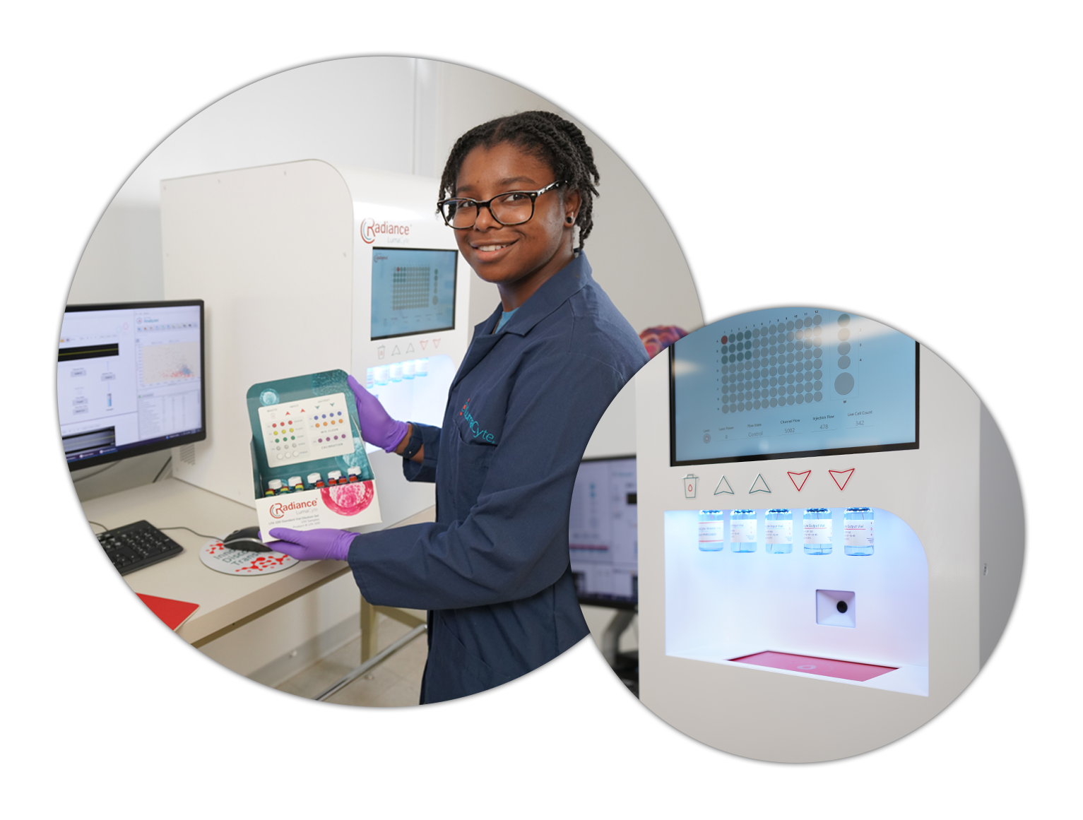
Laser Force Cytology™ (LFC™) measures cellular response and changes to a cell’s intrinsic biochemical and biophysical properties by measuring optical and fluidic forces in a microfluidic platform technology instrument called Radiance®.

An optical force is generated as photons from a laser interact with a cell in a microfluidic channel. The drag force of the fluid acts on the cell in the opposing direction as it flows through the channel.
LFC™ measures velocity (optical force) and other parameters to detect subtle phenotypic changes in cells, rapidly measuring quantitative early indicators of cellular response to viral infection, activation, transfection, transduction and differentiation.

Factors Affecting Optical Force
Subtle phenotypic changes in cell biochemistry and morphology (cytoskeletal changes), which are often associated with cancer, sepsis, and other diseases, give rise to detectable differences in optical force, deformability and a host of other multivariate data parameters quantitated by LumaCyte’s platform technology instrument, Radiance®.

Shape

Surface morphology,
coatings

Refractive index (proteins,
cytoplasm, membrane)

Internal cellular structure
and organization
The Value of Label-Free LFC™ Analysis
Analytical challenges in biomanufacturing impede real-time, high-throughput analyses, limiting the industry’s capacity to optimize processes, improve product quality, and ensure regulatory compliance. Overcoming these hurdles requires the development and adoption of innovative analytical solutions that align with the dynamic nature of the biopharmaceutical landscape.
Antibody Based Assay Limitations
Can induce phenotypic changes in cells, influencing their behavior and characteristics during the staining and labeling processes
Non-specific binding can cause false positives; in other situations, labels are not always uniformly expressed and hence not detected, leading to false negatives
Requires prior knowledge and development, limiting the discovery of unknowns
Label use is error prone, time consuming, and very expensive
Laser Force Cytology™ as a Label-Free Solution
Protects the innate cellular response through a label-free measurement approach
Reduces errors in assay results through unbiased cellular measurements
Enables discoveries of new cells, phenotypes, and cellular biomarkers
Delivers higher quality results in less time and at less cost to the researcher
Subtle cellular changes are precisely captured with Radiance®, allowing developers to measure real-time product quality attributes, rapidly optimize processes, and significantly improve production consistency, yields and shelf life.
Laser Force Cytology™ Compared to Current Analytics
LFC™ provides numerous advantages over current methods for a number of applications, including viral infectivity. The table below provides a comparison between LFC™ and several other common analytical techniques.
|
|
qPCR/ddPCR | Flow Cytometry | Viability | ELISA | TCID50 | |
| Data Quality | High | Medium | High | Low | Low | Low |
| Method of Detection | Cell-Based | Nucleic Acid | Cell-Based | Cell-Based | Protein | Cell-Based |
| Label/Reagent Free | ✓ | X | X | X | X | ✓ |
| Automation | ✓ | ✓ | X | ✓ | ✓ | X |
| Labor Requirements | Low | Medium | High | Low | Medium | High |
| Time to Result (TTR) | Low | Medium | Medium | Low | Medium | High |
| Cost Per Sample | Low | Low/Medium | High | Low | Medium | Medium |
| New Cellular Characterization | ✓ | X | X | X | X | X |
| Real-Time PAT | ✓ | X | X | ✓ | X | X |
Real-Time Process Analytical Technology (PAT) for Continuous Production Monitoring
Advanced bioanalytics play a pivotal role in the success of biomanufacturing by providing crucial insights into process development and ensuring stringent quality control. In biopharmaceutical production, these sophisticated analytical tools enable researchers to monitor and optimize key parameters, such as cell culture conditions, protein expression, and purification processes. The precision afforded by advanced bioanalytics not only enhances the efficiency of biomanufacturing processes but also ensures the consistency and quality of the final product. From accelerating process development to guaranteeing adherence to regulatory standards, these tools are indispensable in the pursuit of robust and reliable biomanufacturing practices, ultimately contributing to the production of safe and effective biotherapeutics.
Laser Force Cytology™ Detects Cellular Changes Unresolved by Flow Cytometry
Below is a set of graphs comparing flow cytometry and LFC™ data of mouse raw macrophages that have been exposed to 5 µm silica beads. In the flow cytometry data, the forward and side scatter of the macrophages with and without silica beads is entirely overlapped and thus undetectable.
